In an ECG pattern the T. The transition from the ST segment to the T-wave should be smooth and not abrupt.
Pin On Ecg
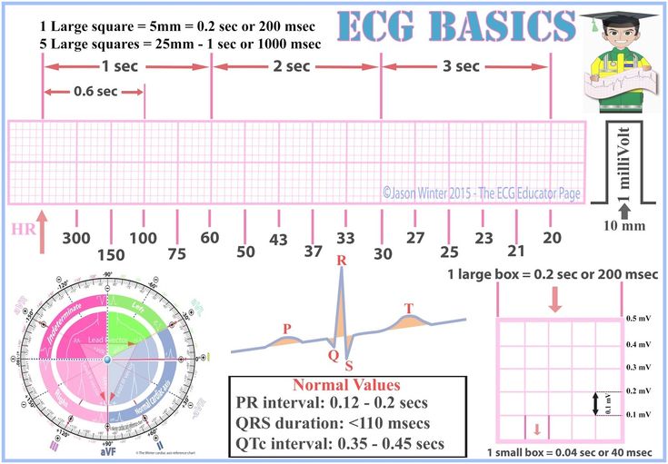
012 to 020 second.

. The T wave on an electrocardiogram ECG represents typically ventricular repolarization. It is positive in all leads except in aVR. The EKG or ECG components are the P wave contraction of the atria the QRS complex the contraction of the ventricles and the T wave repolarization of the ventricles.
The action potential starts at the SA node present in the right atrium and from the atria it passes to the ventricles through the AV node and bundle of His. The T Wave The T wave represents ventricular repolarization. The normal T-wave in adults is positive in most precordial and limb leads.
What part of the ECG tracing represents the time it takes for the impulse to activate the myocardium to the complete contraction. T wave It represents the repolarization of ventricles and the end of the systole. The ventricles are so large that when they contract depolarize the form a large electrical impulse that presents the QRS complex.
Ventricular repolarization What does the P-Q interval represent. This bump is called the t-wave and is caused by the ventricles relaxing. What does the T wave of the electrocardiogram ECG represent.
The first deflection is the P wave associated with right and left atrial depolarization. The T wave on an ECG tracing represents A. Therefore because they are so large when they relax repolarize they form a small electrical impulse that presents as the t-wave.
It follows the QRS complex. Why isnt there a wave on a normal ECG tracing that represents atrial repolarization. Course Title BIOLOGY 2458.
Atrial and ventricular depolarization and repolarization are represented on the ECG as a series of waves. What does the T wave on an ECG tracing represent. Mitral valve prolapse Digoxin effect Right and left ventricular hypertrophy with strain The U Wave.
The T-wave The T-wave reflects the rapid repolarization of contractile cells phase 3 and T-wave changes occur in a wide range of conditions. In leads V1-V4 in children young people and women. However various waveform morphologies may present as an indication of benign or clinically significant injury or insult to the myocardium.
8 At which point on an ECG is the first heart sound LUBB heard. In electrocardiography the T wave represents the repolarization of the ventricles. Possible Causes of T wave changes.
QUESTION 7 The T wave on an ECG tracing represents A atrial repolarization B. 6 What does the T wave represent in an ECG. 5 Which tracing in a normal ECG is the repolarization of the atria.
What part of the ECG tracing represents the repolarization of the bundle of His and Purkinje fibers. In general it has a smaller amplitude than the QRS complex than precedes it. The interval from the beginning of the QRS complex to the apex of the T wave is referred to as the absolute refractory period.
Atrial depolarization Step-by-step solution Step 1 of 4 Electrical impulses which are generated during a cardiac cycle can be recorded by electrocardiograph. Electrical changes are studied using 12 electrodes or leads. The T-wave amplitude is highest in V2V3.
The patients nurse tells you that he has not eaten anything in the past 18 hours. In electrocardiography the T wave represents the repolarization of the ventricles. What factors might lead to differences in the relative magnitude of waveforms.
School University of Texas Arlington. An EKG tracing is a series of boxes upon which positive and negative deflections or waves that represent the electrical impulses of the heart are recorded. The P wave followed by the QRS complex and the T wave.
T-wave changes are frequently misunderstood in clinical practice which the discussion below will attempt to cure. Wave of atrial repolarization is invisible because of low amplitude. Understanding the differential diagnosis for T wave discrepancies is crucial to the successful and safe management of various cardiac.
Electrocardiogram ECG Abnormalities in the QRS complex. The T wave on the ECG T-ECG represents repolarization of the ventricular myocardium. The amplitude diminishes with increasing age.
Its morphology and duration are commonly used to diagnose pathology and assess risk of life-threatening ventricular arrhythmias. Represents time it takes for signal to travel throughout atria. The T wave represents ventricular repolarization.
What does the T wave represent on an ECG. The time between depolarization of S-A node and depolarization of A-V node. 7 Why is QRS complex a downward deflection in human ECG.
Question 7 the t wave on an ecg tracing represents a. A typical ECG tracing of the cardiac cycle heartbeat consists of a P wave atrial depolarization a QRS complex. Each EKG has a baseline or isoelectric line which represents the absence of electrical activity.
Ischemia and myocardial infarction Pericarditis Myocarditis Cardiac contusion Acute neurologic events such as a subarachnoid bleed. The T wave on an ECG tracing represents - ScieMce The T wave on an ECG tracing represents 0 votes More questions like this You receive a call to a skilled nursing facility for an elderly man with generalized weakness. The T wave on an ECG tracing represents A atrial depolarization.
Pages 20 Ratings 98 56 55 out of 56 people found this document helpful. Each of the following changes will result in increased blood. Generally the T wave exhibits a positive deflection.
The reason for this is that the last cells to depolarize in the ventricles are the first to. T Wave It represents ventricular repolarisation. It could be negative in lead III in obese patients.
EKG Chapter 2 - cardio system. Physiology variants and ECG features The normal T-wave Assessment of the T-wave represents a difficult but fundamental part of ECG interpretation. What might a longer-than-normal P-R interval indicate.
BC atrial repolarization happens at the same time as ventricular repolarization during the QRS Complex and ventricular repolarization covers it. The PR interval is usually.

The Heart S Electrical Activity Is Represented On The Monitor Or Ecg Tracing By Three Basic Waveforms The P Wa Nursing Notes Pr Interval Basic

Pin By Abhisheksensual On Tigg A Basic Pr Interval Medical
Ecg Nomal Waves And Segments Medstudent Ekg Basics Waves Segments Definitions Ecgeducator Segmentation Pr Interval Ekg


0 Comments